Component of circulatory system and why is important to the brain, and fetal on pregnancy mother.
BLOOD VESSEL.
Blood vessels is closed tubular network responsible for transport of blood from the heart to the body and to bring it back. These vessels which carry blood away from the heart are called arteries and arterioles. These vessels which carry blood back to the heart are called venule's and veins. Capillaries are microscopic vessels which join the arteries and veins.
Histology of blood vessels.
Blood vessels except capillaries are made up of three distinct layers called tunics.
Three distinct layers are:
1. Tunica internal which is inner layer made by epithelial lining.
2. Tunica media which are middle lining made by smooth muscle and elastic connective tissues.
3. Tunica external adventitia which are out lining made by connective tissues covering.
The thickness and histologic composition of these three layers differs in the arteries and veins which give them their unique structure which enable them perform their unique functions.
The closed space inside the muscular tube is known as lumen.
I. Tunica internal: Tunica internal is innermost layer of blood vessels
It is composed by three layers:
1. Endometrium
2. Basement membrane.
3. Elastic layer
A. Endometrium.
This layer is direct contact with lumen which are:
1. Made up of squamous epithelium.
2. It is surrounds the whole of the cardiovascular system
3. It is smooth surface facilitated the friction less movement of the blood.
4. It releases biochemical which effect the contractile state of vessel.
5. Have role of platelet aggregation and inflammatory processes.
B. The basement membrane.
1. Present deep to endometrium.
2. Give supportive base to the endometrium.
3. Anchors endothelium to connective tissues.
4. Has a frame work of collagen fibers which give it flexibility and tensile strength.
C. Elastic layer
1. Made up of elastic fibers
2. Make boundary between tunica media
II. Tunica Media( Middle layer).
Compose of smooth muscles and elastic fibers display greatest variation in size and composition among different types of blood vessels. The smooth muscles in tunica media media regulate the diameter of vessels.
The smooth muscles in tunica media regulate the diameter of vessels. It is innervated by sympathetic nervous system which media regulate the diameter( vasoconstriction). Play important in blood flow and blood pressure regulation by vasoconstriction and vasodilation, on it outer side in the layer of elastic fiber which separate it from tunica externa.
II. Tunica externa: is the outer covering of the blood vessels, it is made by loose connective tissues.
It plays three important roles which are:
1. Contain the capillaries known as vasa vasorum
2. Which supply the large vessel wall with blood.
3. Cary nerve supply to vessel
III. Tunica externa: is the outer covering of blood vessels it is made by loose connective tissue.
It plays three important role which are:
1. contain capillaries known as vasa vasorum
2. which supply the large vessel wall with blood
3. Cary nerve supply to vessel
4. Anchors the blood vessels to adjoining tissues.
Difference between classification of artery.
|
Component |
Elastic |
Muscular |
Small
artery |
|
Size |
Largest in diameter | Medium in size |
small in diameter (15-300micrometer) |
Composition |
have high elastic fiber in elastic laminas |
have the capacity of vasoconstriction and vasodilation because of smooth muscle |
loose connective tissue with sympathetic nerve supply |
|
Connection |
conduct blood to muscular arteries |
connect with capillary | |
|
Elasticity |
can expand and recoil |
maintain state of constriction known as the vascular tone |
|
|
Function |
function as pressure reservoir |
help in blood flow |
Regulate blood pressure by alternate the resistance |
|
Example | Aorta, pulmonary trunk, subclavian artery |
bronchial artery, femoral artery |
Capillaries
Capillaries is the function unity of cardiovascular system, small in size has 8 to 10 micrometer and is made by endometrium tissues only. Each capillary bed is is supplied by meta arteriole. Meta arteriole regulate blood pressure supply to capillary and also distinct end of meta arteriole has no smooth muscle and is called through fare channel.
Types of arteries
There three types based on structure
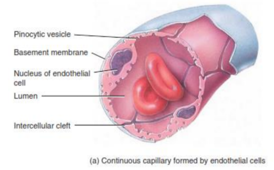
1. Continuous Capillaries
o It allow the exchange of small molecules and other exchange take place by pinocytosis
o More impermeable in the brain it found in the brain, muscle and skin
o Most abundant make continuous tube where cell heled together by the tight junctions.
2.
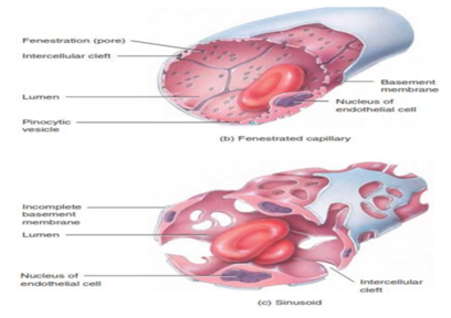
2. Fenestrated capillaries
o In these capillaries the plasma membrane of the epithelium cell has small pores of about 70-10nm called Fenestration. Allow the exchange of slightly layer molecule but large protein cannot pass through it.
o Discontinuous capillaries
Vein
Veins are blood vessels that carry blood to words the heart. Most veins carry deoxygenated blood( Blood that has given up its oxygen to the body's tissues). The exception is the Pulmonary veins which carry oxygenated blood from the lungs back to the heart.
Structure.
1. Veins have thinner walls than arteries
2. The often contain Valves that prevent blood following backwards. .
3. The wall have three layers
Function
A. Return blood to the heart: Veins carry blood back to the heart after it has circulated through the body.
B. Blood reservoir: Veins can hold a large amount of blood, acting as a reservoir.
1. Vascular sinus: A dilated blood vessel, often found in certain organs like the brain or liver.
2. Portal Vein: an enlarged, twisted vein, often found in the legs.
3. Varicose Vein: an enlarged, twisted vein, often found in the legs.
5. Hemorrhoids: Swollen veins in the anus or rectum.
6. Esophageal Vein: A vein located in the esophagus, the tube connects the throat to the stomach.
Arterial system
The Arterial system is a complex network of blood vessels that carry oxygen- rich blood away from the parts of the body.
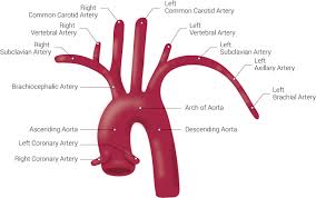
Aorta: the largest artery in the body, branching off the left ventricle of the heart. it further divides into smaller arteries to supply blood to different regions.
Arteries: These are muscular tube that carry oxygenated blood away from the heart.
Arterioles: smaller branches of arteries that further divide into capillaries.
Capillaries: Tiny blood vessels that form a network, allowing for exchange of oxygen, nutrients, and waste products between blood and tissues.
Systematic Circulation
Systematic circulation is the part of circulatory system that carries oxygenated blood away from the heart to the body and returns deoxygenated blood back to the heart. Systematic arteries arise from Aorta.
Aorta is the largest artery of the body with diameter of 2-3 cm. it merge from left ventricle posterior to pulmonary trunk.
On the basis of anatomic location, it can be divided into four segments:
1. Ascending aorta
2. The arch of aorta
3. The thoracic aorta
4. Abdominal aorta
A. Ascending aorta
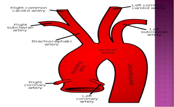
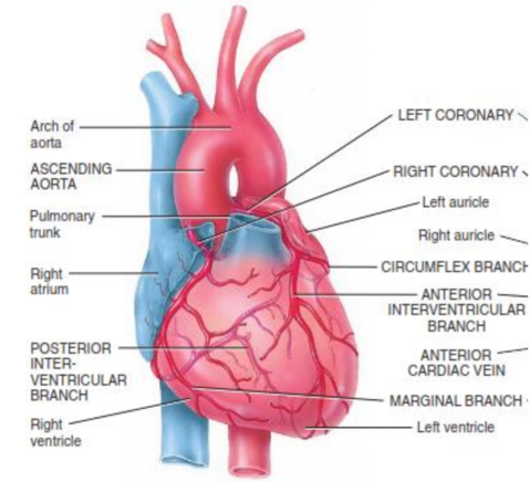
Abdominal aorta The ascending aorta is the first part of the aorta, the largest in human body. it originates from the left ventricle of the heart and ascends slightly to the right of the midline before curving to form the aortic arch. The ascending aorta starts at aortic valve, where it connects to the left ventricle of the heart. it end at the beginning of the aortic arch. Diameter: the diameter of the ascending aorta varies depend on factors like age, sex, and overall health. Normal diameter is less than 4 cm. the Ascending Aorta is about 5 cm in the length and begin in the aortic valves. it covered by pericardium. The ascending aorta give off two branches which are right coronary artery and left coronary artery. Coronary artery divides to form coronary circulation.
It is continuation of aorta. emerge from pericardium posterior to the sternum starts at the moves superior and posterior and turn inferior ( making an arch), ending at the intervertebral disc between 4th and 5th vertebral where it become thoracic aorta.
Three main arteries emerge from the arch of aorta.
- The brachiocephalic artery
- The left common carotid artery
- The left subclavian artery
The brachiocephalic artery
It is the first and largest branch from the aorta, arise from the superior aspect of the arch of aorta moves superior to the right and at the sternoclavicular joint divided into two branches.
A. subclavian artery
Extend from brachiocephalic artery to inferior border of the first rib, give off following branches at the base of the neck.
1. Internal thoracic artery arise from first part of subclavian artery descends posterior to costal cartilages of superior six ribs terminates at sixth intercostal space sending branches into intercostal spaces.
2. Vertebral artery: a major branch to the brain.
3. Thro-cervical artery: a short trunk serving thyroid gland, trachea shoulder and larynx.
4. Costocervical artery: serve the upper intercoastal muscles , posterior neck muscles and spinal cord and its meninges.
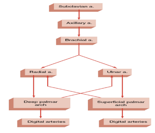
B. Axillary artery
After give off branch in thoracic cavity, the artery enters the axillary region at the inferior border of first rib. It gives off numerous at the axilla supplying blood tot he thorax and shoulder region. it end at the distal end of teres major muscles.
1. Branchial artery.
It is the continuation of axillary artery to the arm ( subclavian, Axillary and branchial artery in the same artery with different name at different locations) Superficial and palpable at the media side of the arm as it descends through the elbow joint it curves laterally passing through cubital fossa where it is easily palpable.
It supplies blood to muscles of arm, hummers and elbow joint, gives off deep branchial artery ( supplies triceps branchial) and anterior and posterior humeral circumflex arteries( forms anastomotic network around hummers)
Terminates by dividing into proximal to cubital fossa
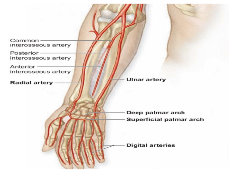
Arteries of the fore arm: just proximal to the cubital fossa, (a triangular depression that lies in front of the elbow) the branchial artery branches into the radial and ulnar arteries, which supply blood to forearm, hand and digits.
I. Radial artery.
Radial artery courses down the lateral, or radial, side of the arm, where it send numerous small branches to the muscles of the forearm. Branches of radial arteries: The Radial recurrent artery serves the region of the elbow and the first and largest branch of radial artery. the radial artery divides into deep radial artery and superficial redial artery at the wrist.
II. ulna artery.
Large among the 2 from branchial artery the ulnar artery extends down the medial, or ulnar, side of the forearm and gives off many small branches to the muscles on that side.
Branches of Ulnar Arteries
o Ulnar Recurrent artery, which arises from the proximal portion near the elbow.
2. common interosseous artery which divides into anterior and posterior interosseous arteries and at wrist it branches into superficial and deep branches that enter hand, the radial and ulnar arteries anastomose giving rise to coronary arteries (CA).
Superficial palmer arch
Formed mainly by superficial palmar branch of ulnar artery with contribution from superficial branch of radial artery, present superficially, gives off digital arteries to the fingers. Deep palmer arch
Arise mainly from deep palmar branch of radial artery contribution from deep branch from ulnar artery, present deep in palm, give rise to metacarpal arteries.
o The common carotid arteries course upward in the neck along the lateral side of the trachea.
o Right common carotid artery merges from bifurcation of the brachiocephalic trunk while the left common carotid artery emerges directly from the arch of aorta.
o Each common carotid artery branches into the internal and external carotid arteries slightly below the angle of the mandible.
o At the base of the internal carotid artery is slightly dilation called the carotid sinus. The Carotid contain baroreceptors, which monitor blood pressure.
o Surrounding the carotid sinus are the carotid bodies, small neurovascular organs contain chemoreceptors, which respond to chemical changes in the blood.
o The internal carotid artery ascends in the neck until it reaches the base of skull, where it enters the carotid canal of the temporal bone.
The internal carotid artery gives off branches during it course. the important among which are:
- Ophthalmic artery: supply eye and associated structures.
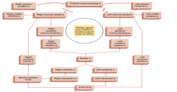
- Cerebral artery( Anterior and middle): Supplies the cerebrum. The paired internal carotid arteries along with paired vertebral arteries( subclavian arteries) make the four arteries that supply brain. At the inferior surface of the brain around the pituitary gland the branches of these vessels anastomose making at the cerebral arterial circle.
The circle of willis
The circle of willis is formed by two vertebral arteries and two carotid arteries, arise from the subclavian artery at the base of neck superiorly through the cervical vertebrae and enter the skull through the foramen magnum. in the cranium (at the level of pons) they unite to form basilar artery. The basilar artery ascend along the inferior surface of the brain stem and terminates by forming that supply the portion of cerebrum.
- On the anterior side the internal carotid arteries give off cerebral arteries anastomose with each other though anterior communicating artery.
- On the posterior side the the internal carotid arteries anastomose with the posterior communicating arteries.
The anastomoses of vertebral arteries making basilar artery, the anastomoses at anterior communicating artery and anastomoses of internal carotid arteries with posterior, communicating arteries making collective make the circle of willis.
External Carotid Artery.
Major blood sources to all structures of the head except brain. External carotid Artery gives off several branches as it extends upward along the side the of the neck and head. these are:
1. Superior thyroid artery: Which serves the larynx and vocal colds and the thyroid gland.
2. lingual artery : Which provide extensive vascularization to the tongue and sublingual gland.
3. Facial artery: Which traverses on inferior margin of the mandible to serve the pharyngeal area, palate, chin, lips, and nasal region.
4. Occipital artery: Which serves the posterior portion of the scalp, the meninges over the brain and certain posterior neck muscles.
5. Posterior auricular artery: Which serves the auricle of the ear and the scalp over the auricle. The external carotid artery terminates at a level near the mandibular condyle by dividing into maxillary artery and superficial temporal artery
- The maxillary artery give off branches to the teeth and gums, the muscles of mastication, the nasal cavity, the eyelids and the meninges.
- The superficial temporal artery supplies blood to the parotid gland and to the superficial structures on the side of the head.
Thoracic Aorta
It has 20 cm long, continuation of aorta, its begins at the at the level of the intervertebral disc between the fourth and fifth thoracic vertebrae, where it lies to the left of vertebral column. as it descends, it moves closer to the midline and extends through an opening in the diaphragm called aortic hiatus at the level of the intervertebral disc between the twelfth thoracic and first lumbar vertebrae. During its course the thoracic aorta gives off numerous branches to viscera (Visceral branches) and wall structures (parietal branches).
Branch of Thoracic Aorta
A. Visceral branches
1. Pericardial artery: Arise at various levels of aorta and move forward to supply the pericardial sac.
2. Bronchial artery: Arise either directly from aorta or from any of its branch supplies the bronchial.
3. Esophageal artery: arise from anterior surface of aorta and supplies all of the esophagus.
4. Mediastinal arteries: Arise from various levels of mediastinum and supplies the supplies the mediastinal connective tissues and lymph nodes.
B. Parietal branches
Nine pairs of arteries that arise from posterolateral aspect on each side of thoracic aorta . Each passes laterally and then anteriorly through intercostal space, where they will eventually anastomose with anterior branches from internal thoracic arteries. supplies the muscles and ribs of thoracic wall, meninges and spinal cord arise from lower part of thoracic aorta, extends to lower thoracic wall and upper abdominal body wall.
Supplies the skin muscles and lower thorax and spinal cord meninges arise the lower part of thoracic aorta extends to the superior surface of diaphragm supplying the diaphragm muscle and pleura.
Abdominal Aorta
The abdominal portion of the aorta is the segment of the aorta . where it divides into the right and left common iliac arteries. The abdominal aorta lies anterior to vertebral column. It gives off both parietal and visceral branches.
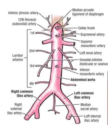
I. visceral Branches
Unpaired
- Celiac trunk
- Superior Mesenteric Artery
- Inferior Mesenteric artery
Paired
- Suprarenal arteries
- Renal arteries
- Gonadal arteries
II. Parietal Branches
Paired
- Inferior phrenic arteries
- Lumbar arteries
Unpaired
- Median sacral artery
I. Unpaired Visceral branches
Celiac trunk arise from the ventral aspect of aorta the esophagus( Abdominal component), stomach, duodenum and spleen. it give off five branches which are:
1. Left gastric arteries supplying the esophagus and stomach.
2. Splenic artery that gives off branches to spleen, pancreas and stomach (pancreatic, left gastroepiploic, short gastric).
3. Common hepatic artery that gives branches to liver, gallbladder, pancreas and stomach(proper hepatic, right gastric, gastroduodenal).
4. Superior mesenteric artery arise from the ventral surface of abdominal aorta inferior to celiac artery. Supplies jejunum, ileum, and ileac, ileocolic, right and middle colic have extensive anastomoses
5. Superior mesenteric artery: arise from ventral aspect of aorta inferior to superior mesenteric and supplies the large intestinal from transverse colon to rectum, through its main three branches( left colic, sigmoid and rectal)
II. Paired Visceral branches
1. supra renal artery: arise from the lateral aspect of aorta. Mostly three arteries arise from aorta other may rise from other branches of abdominal aorta, supplies the suprarenal glands (adrenal grand)
2. Renal artery: arise from lateral aspect of abdominal aorta supplies the kidneys
3. Gonadal artery : arise from lateral aspect of aorta just inferior to renal arteries. in male they are called testicular passes down the abdominal wall reaching the scrotum. In female they are called ovarian arteries.
III. Paired parietal branches.
1. Inferior phrenic artery: arise from lateral aspect of aorta superior to celiac trunk, supplies the diaphragm.
2. Lumbar artery: arise from dorsal (posterolateral) aspect of aorta supplies the lumbar vertebrae, spinal cord and muscles of abdominal wall.
IV. Unpaired Parietal Branches
1. Median sacral artery: arise from the dorsal aspect of aorta supplies the sacral and coccyx vertebrae and spinal nerves.
2. Lumbar artery: at the posterior pelvic area the abdominal aorta bifurcates to right and left iliac arteries.
3. Iliac arteries: the iliac arteries go down the pelvic and terminates by dividing into internal and external iliac arteries.
the internal iliac arteries branch extensively and supplies the gluteal muscles and pelvic organs( Important branches are gluteal iliolumbar, gluteal, vesicular uterine and vaginal, pudendal arteries). The External artery pass out of pelvic cavity to the inguinal ligamental and become femoral arteries.
Femoral artery
The femoral artery passes through the where it become superficial and it palpated. it downwards and posterior to knee where it is called popliteal artery
1. The popliteal artery gives off small branches to knee joint and then divides to anterior tibial artery and posterior tibial artery.
2. the anterior tibial artery runs on the anterior aspect of leg supplying the foot muscles . At ankle it becomes which supplies the ankle and dorsum. it can be palpated there an is the distal pulse of the body.
3. The posterior tibia artery runs on the posterior aspect of leg, at the ankle it divides into lateral plantar artery and medial plantar artery where, the lateral plantar anastomoses with dorsal pedal artery making plantar arch which gives off digital arteries to the toes.