What are difference between Anatomy of Female Pelvic and male pelvis?
The female pelvis is structurally adapted for child bearing and delivery.
There are four pelvic bones
1. innominate or hip bones
2. Sacrum
3. Coccyx
A. Innominate bones
Each innominate bone is composed of three parts.
1. The ilium the large flared out part.
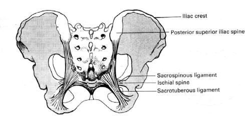
2. The ischium the thick lower part. It has a large prominence known as the ischial tuberosity on which the body rests when sitting. Behind and a little above the tuberosity is an inward projection, the ischial spine. In labor the station of the fetal head is estimated in relation to ischial spines.
3. The pubis The pubic bone forms the anterior part. The space enclosed by the body of the pubic bone the rami and the ischium is called the obturator foramen.
B. The sacrum
The Sacrum is a wedge shaped bone consisting of five fused vertebrae. The upper border of the first sacral vertebra is known as the sacral promontory.
The anterior surface of the sacrum is concave and is referred to as the hallow of the sacrum.
C. The coccyx
The coccyx is a vestigial tail. It consists of four fused vertebrae forming a small triangular bone.
Pelvic Joints.
There are four pelvic joints
- One Symphysis pubis
- Two Sacro iliac joint
- One Sacro coccygeal joint
- The symphysis pubis is a cartilaginous joint formed by junction of the two pubic bones along the midline.
The Sacro iliac joints are the strongest joints in the body.
- The Sacro coccygeal joint is formed where the base of the coccyx articulates with the tip of the sacrum. In non pregnant state there is very little movement in these joints but during pregnancy endocrine activity causes the ligaments to soften which allows the joints to give & provide more room for the fetal head as it passes through the pelvis.
Pelvic ligaments
Each of the pelvic joints is held together by ligaments
- Inter-pubic ligaments at the symphysis pubis (1)
- Sacro iliac ligaments (2)
- Sacro coccygeal ligaments (1)
- Sacro tuberous ligament (2)
- Sacro spinous ligament (2)
The True Pelvis
The true pelvis is the bony canal through which the fetus must pass during birth. It has a brim, mid cavity and an out let. The pelvic brim is rounded except where the sacral promontory projects into it.
The pelvic cavity is extends from the brim above to the out let below.
The pelvic out let are two and described as the anatomical and the obstetrical.
- The anatomical out let is formed by the lower borders of each of the bones together with the Sacro tuberous ligament. It is diamond in shape.
- The obstetrical out let is of the space between the narrow pelvic strait and the anatomical outlet. Important land marks of female pelvis
A. Pelvic brim
- Sacral promontory posteriorly
- Superior ramus of the pubic bone antra lateral
- Upper inner boarder of the body of the pubic bone
- Upper inner boarder of the symphysis pubis anteriorly
B. Mid pelvis
- Ischial spine
C. Out let
- Inferior pubic rami antero laterally
- Sacro tuberous ligament postro laterally
- Ischial tuberosity laterally
- Inferior border of symphysis pubis anteriorly.
- Tip of coccyx
Important diameters of the pelvis
I. Inlet
Diagonal conjugate - a line from the sacral promontory toward the lower boarder of the symphysis pubis and measures 12.5 centimeter.
It is measured by pelvic examination.
II. Mid cavity
Interspinous diameter a line between the two ischial spines and measures 11 centimeter.
III. The pelvic out let
- Pubic arch
- Inter-tuberous diameter
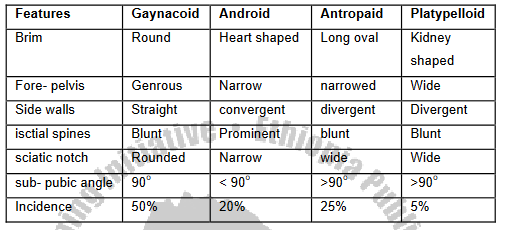
The four types of female pelvis
1. The gynecoid pelvis (female type)
2. The android pelvis (male type)
3. The anthropoid pelvis
4. The platypelloid pelvis
After Reading this article test yourself on our Quiz Part and Goodluck!